![]()

![]()
|
|
Getting Started with UltrasoundContents: Ultrasound BasicsUnderstanding Sonographic Pictures Diagnostic Ultrasound & Podiatric Pathology Sound-Seal™ Protective Film Dressing Wound-Mapping® Ultrasonic Assessment Method INTRODUCTIONUltrasound (sonography) is a medical diagnostic technique which allows one to visualize and therefore examine a part of the human anatomy. The process incorporates the use of high frequency sound waves emitted from a probe and directed into a body. These sound waves penetrate and encounter the different tissue interfaces as they travels through the body. When sound encounters tissues or tissue planes, part of the wave is reflected back to receivers in this same probe. Different tissue interfaces cause a "reflective pattern" which is then sent to a computer. The computer processes the information and produces an image, which is sent to a video screen, printer, and or videocassette recorder. These images are then studied either "live" or at a later time to rule out any number of pathologies. Ultrasound waves are produced by oscillating crystals at a frequency that is inaudible to the human ear. Transducers located in the probe produce sound (for example) at 7.5mhz which is then pulsed at intervals which occur every 20 micro-seconds. These same transducers after transmitting the sound then capture the reflected echoes. The transducer must be in contact with the medium scanned, in this case skin, so a "transmission jelly" is used to insure a complete "union". The ultrasound produced can not travel through the air and then into the body. Ultrasonography Imaging We are all familiar with Doppler studies which is the evaluation of the "flow" of a substance such as blood through a vein or an artery. However the ultrasound imaging that we are discussing here is either the B-mode or M-mode. Ultrasound (sonar) has its roots in the military, and in brief, it was used in submarines for detecting the surface and shape of the ocean floor. In medicine, B-mode or B-scan simply means the scanning of a part of the human body. Such a scan is either live, where the image is seen on a video screen and printed at the time it is acquired, or scanned now and compiled later into and image for study. (The later was an earlier method to produce an image for study.) This is 1998 CPT code # 76880- Echography, extremity, non-vascular, B-scan and/or real time with image documentation. M-mode indicates that there is the recording of MOTION, such as that of an ultrasound of the heart or a fetus. Briefly, if one takes a scan and produces a picture of a fetus within the mother, we could not tell if it was alive at that moment. However, a B-mode image along with an M-mode image recorded at that same time will show that the fetus was alive at that point in time. As ultrasound waves are sent into the tissues, part of the sound is reflected back to the probe while others continue into deeper tissues. Much of this is dependent on the frequency of the probe, which is being used. Unlike the probes used for physical therapy which are designed for deep heating of tissues, diagnostic ultrasonography probes cause no heating or tissue damage. For diagnostic musculoskeletal ultrasound a 7.5mhz probe is used. This 7.5mhz probe produces an image that is 4.5cm long and penetrates into the tissues 7cm deep at maximum. This 7.5mhz probe known as a "high resolution extremities probe" is used for carpal tunnel syndrome, torn rotator cuff, and has many excellent podiatric applications. Other probes such as the 5.0mhz are for deeper structures (used for the examination hip, knee, and spine), and 3.0mhz for still deeper structures. Ultrasound can be used to examine the arterial/venous system, heart, pancreas, urinary system, ovaries, spinal cord, and more. Ultrasound is excellent for diagnosing cysts in soft tissue because usually they contain fluid. Ultrasound is safe during pregnancy. Ultrasound uses no radiation unlike x-rays, Cat Scans and other such modalities. UNDERSTANDING Sonography PICTURES When the Ultrasound wave encounters a dense object such as bone, most of the sound is bounced back up to the probe and little is allowed to pass through the tissue. This is seen as bright white on the video screen, and known as HYPERECHOIC very ECHOGENIC. In contrast if one scans a ganglion cyst, (a fluid filled sac) the video display reveals an oval dark area within tissues. This is known as HYPOECHOIC and ANECHOIC. The position of structures within the scanned field are described as NEAR FIELD (anterior) and FAR FIELD (posterior). Further, structures within the scanned field are described as HOMOGENEOUS or uniform in pattern, and HETEROGENEOUS or irregular in pattern. For most, the first contact and initial examination performed by one not familiar with diagnostic ultrasound will require some study. We would like to see a picture such as that of an MRI or CAT scan. However, the physician quickly realizes that the video or picture that is being studied is a small section of tissue. Examples of Ultrasound scans like those that you will encounter are found in the Case Study and Other Scans pages on this web site. Scanning Podiatric PathologyThe position of the probe and its anatomical location is extremely important. Unlike that of x-rays, many of the longitudinal or transverse scans may initially look the same. It is essential that the physician becomes familiar with the different "tissue pattern" (e.g. a tendon Vs bone Vs muscle etc.). The way a particular type of tissue responses to an ultrasound wave must be learned. Once the normal structure is recognized, the diagnosis of pathology becomes apparent. With this in mind bilateral examinations are common. Scanning the opposite limb (or the normal limb) for a patient is very helpful. An advantage of diagnostic ultrasound besides its safety, is the ability to perform live active and/or passive range of motion studies. The ability to move a particular part of the anatomy, observe and record motion studies such as partial or complete tendon, muscle, and ligament tears should not be understated. To enable the physician to diagnose cases, record, and document these studies increases the likelihood of better treatment and end result. Please note that MRI, CAT scans, and x-rays all do not allow for motion studies. Although diagnostic ultrasound is not intended to eliminate those studies, one should realize that the cost of an ultrasound is about one tenth that of an MRI. Should the physician receive satisfactory information from the musculoskeletal ultrasound and avoid the need of an MRI scan, the patient and/or insurance companies benefit in those savings. DIAGNOSTIC ULTRASONOGRAPHY AND The foot, perhaps more than any other area of the body receives a lot of injury, stress, and strain. The reaction of tissue to trauma is the same throughout the body causing pain, swelling and the like. Trauma and resulting swelling and inflammation cause an influx of fluid to a particular area. In diagnostic musculoskeletal ultrasound, scanned areas of inflammation appear darker or hypoechoic due to the sound wave passing through the fluid filled inflamed area. Pathology in which ultrasound will aid the podiatric physician in the office setting includes:
As stated earlier ultrasound studies require the physician to place the probe on an area in the transverse (frontal) plane or the longitudinal (sagittal) plane. Knowledge anatomical structures and their relative position is a must (i.e. what is superior, inferior, proximal, or distal).
|
Image Library Ultrasound Images of the foot & ankle
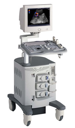
Looking For an Ultrasound Scanner? Click Here
Image Library Ultrasound Images of the foot & ankle
Image Library Ultrasound Images of the foot & ankle
Image Library Ultrasound Images of the foot & ankle
Image Library Ultrasound Images of the foot & ankle
|

| Copyright © 2004 -
BioVisual Technologies LLC-
|